Branka Krsti}
Faculty of Agriculture, University of Belgrade, Beograd-Zemun, 11080,
Yugoslavia
The nature, the origin, the architecture and the replication of viruses represent significant biological questions, not only for viruses themselves, but also for other living forms because learning about them contributes to better understanding of the evolution of the living world.
The evolution of viruses in nature is performed in two levels. Micro-evolution means smaller changes of genome by means of the substitution, addition or deletion of nucleotides and leads to the creation of strains and other forms within the species. Macroevolution leads to sudden major changes by duplication of existing genes, by recombination with related viruses or by the acquisition of a gene from the host or an unrelated virus, which lead to the creation of new species (Matthews, 1992). By these ways of evolutionary development, viruses, as spiritless wonders of nature, have brought to the perfection their forms, architecture and replication mechanisms by using different and always efficient ways and means for the realization of the purpose of their existence - to produce infectious progeny.
The basic structural requirement for plant viruses is the provision of a shell of protein to protect its nucleic acid. Their capsid consists of structurally and functionally identical protein subunits that are repeated in the particle by binding in the regular manner using a geometrical plan and a certain type of symmetry. Viruses have adapted their structure ti their bilogical functions forming particles of uniform size, shape and composition. The basic rule in forming virus particles is established on limited genetic information. Because of such genetic information, viruses have a large number of identical protein subunits which have instructions for self-assembling. These identical units are packed in a regular manner by specific binding and, in this way, well organized symmetrical structures are created (Kaspar, 1970, Matthews, 1992, [uti}, 1994).
During their evolutionary development, by the repetition of their structural elements, viruses have managed to develop perfect structures of certain space-arrangement and symmetry, with the maximum number of stable mutual bonds, so that such structures are with the minimum free energy. Such stable structures enable an infectious nucleic acid to be preserved, i. e. viral genome is safely packed in them until it comes to a cell, where it could only function.
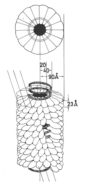
Fig 1. Drawing of the intact particle
of tobacco mosaic Tobamovirus - Helical arrangement of the subunits
in relation to the RNA in a tobacco mosaic virus particle. Each protein
subunit is shaped roughly like a shoe. In successive turns of the helix,
the subunits form a groove in which the RNA is situated (Nienhaus, 1985).
The basic life-process of viruses, multiplication, which means infectivity and parasitism at the same time, and consequently leads to chaos in an infected cells, is also brought to the perfection. How otherwise to explain the process which happens at level of the most responsible cellular functions, by using cellular energy, cellular components, the mechanisms of the synthesis of proteins and nucleic acids and, on the other hand, leads to the collapse of a cell itself. And how to explain that viruses, with limited genetic information and without their own metabolism, can direct such a perfect machinery as a cell to the synthesis of its own parts. This process has been brought to the perfection probably in order to enable the existence of viruses as a species.
Such a perfect process as the multiplication of viruses, makes chaos in an infected cell, which is manifested in the derangement of its structure and function, and which results in various cellular deformities and disintegration. Viruses have brought all the processes necessary for their own existence into such harmony that making chaos in the cells of attacked organisms, they do not annihilate and destroy themselves. By this chaos viruses produce infectious progeny and prolong their biological function.
In contrast to that, man has come to chaos, in which we are today,
by loosing the whole and the harmony of living, considering the question
of purpose unnecessary. All of that causes the turning of sense into madness,
absurdity and destruction in which we are also situated nowadays.
Viruses do not divide and do not produce any kind of specialized
reproductive structures. Instead, they multiply by inducing host cells
to form more viruses. Viruses do not cause disease by consuming cells or
killing them with toxins, but by utilizing cellular substances during multiplication,
taking up space in cells, and disrupting cellular processes (Agrios, 1997).
As metabolically inert and energetically dependant, viruses have found the unique way of their replication. Although many aspects of their replication in vivo (for example - the sites, time and mechanisms by which infecting viral genomes become uncoated, the mechanisms by which an infecting nucleic acid organizes characteristic replication sites within the cell, the mechanisms of modification of cell's organelles, etc.) have already been brought to light so far, there is still the question of how viruses initiate a cell to form as much as possible new particles by itself, coming to its own chaos during that.
And what has been known by now? Although of simple chemical structure, viruses are characterized by diverse nature of their nucleic acid. Expect the viruses with ssRNA (+), those with ssRNA (-), ssDNA and dsDNA are also discovered (Matthews, 1992). Different kind of viruses replicate in different ways, depending on the structure of viral genome, but the process is basically the same.
The replication of viruses is performed through several phases. Animal viruses have the phase of adsorption of particles on the surface of a cell by the certain receptors. At this level, a virus and a susceptible animal cells recognize each other. There is no recognition at that level with plant viruses, which is determined by differences in the build of a plant and animal cell. Plant viruses enter cells only through wounds which are made mechanically or by vectors, or by deposition into an ovule by an infected pollen grain. During the simplified replication of an RNA virus, its nucleic acid firstly gets rid of the protein coat and in that way the viral genome becomes free. By brining the viral genome into a cell, the genetic determinants which code the synthesis of macromolecules specific to a virus, are also brought. The redirection of the cell's metabolism is performed by early proteins-enzymes (polymerase) which are coded by the viral genome. It is thought that one of the ways of the recognition of plant viruses and a susceptible host plant is exactly the synthesis of proteins which are the components of polymerase. The ability of a virus to control a cell's metabolism depends on the nature of the virus and on the type of the cell. At the phase of biosynthesis viruses transmit the genetic information in different ways, and it is basic that specific mRNA must be transcribed (transcription) from the viral genome. When it is once achieved, viruses then translate (translation) mRNA into structural and non-structural proteins by using cellular components and mechanisms. After this phase, proteins and nucleic acid are self-assembled into viral particles, which are further able to infect.
During the self-assembling of the identical units one or more ordered structures could be created. Which of them will be chosen for the plant virus structures depends on its stability and on which of them uses least energy for its building. For plant viruses it has been established that they are either tubes with helical or closed shells with icosahedral symmetry. These structures offer an efficient way for packing viral nucleic acid.
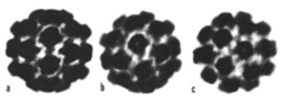
Fig 2. Surface features revealed by negative staining in an icosahedral turnip yellow mosaic Tymovirus with 180 subunits clustered into 12 pentamers and 20 hexamers (Matthews, 1992).
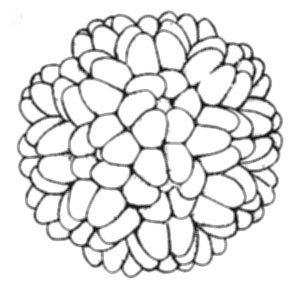
Fig 3. Drawing of the outside of the particle of turnip yellow mosaic Tymovirus showing clustering of protein subunits into groups of five and six (Horne, 1974).
Normally, plants develop different ways of resistance to viruses as to agents which bring them danger. But nature has made it possible that viruses overcome this obstruction by their great and relatively quick variability.
At the end, there is a question which could be asked - Why doesn't
man, as the only being gifted with spirit, learn from some simpler forms
of life, such as viruses, and how could he establish harmony in his surroundings
to survive?
LITERATURE
Agrios, G. N. (1997): Plant Pathology. Academic Press, San Diego, London,
Boston, New York Sydney, Tokyo, Toronto.
Kaspar, D. L. D. (1970): Shematski principi u gra|i virusnih ~estica.
U: Horsfall, F. L. Tam, I. (eds.): Virusne i rikecijske infekcije ~oveka
(Viral and Rickettsial Infections of Man ). Vuk
Karad`i}, Beograd.
Matthews, R. E. F. (1992): Fundamentals of Plant Virology. Academic
Press, Inc., San Diego, New York, Boston, London, Sydney, Tokyo, Toronto.
[uti}, D. (1994) Biljni virusi
(Plant Viruses). Poljoprivredni fakultet, Beograd-Zemun.
Horne, R. W. (1974): Virus structure. Academic Press, Inc. New York,
London.
Nienhaus, F. (1985): Viren, Mykoplasmen und Rikettsien-parasiten an
der Schwelle des Lebendigen. Verlag Eugen Ulmer. Stuttgart.
 Branka
Krsti}, Ph. D., is an Associate Proffesor of Plant Virology and
Anatomy and Physiology of Diseased Plants at the Institute of Plant and
Food Protection, Faculty of Agriculture, University of Belgrade, Belgrade-Zemun,
Nemanjina 6, 11080 Zemun, Yugoslavia.
Branka
Krsti}, Ph. D., is an Associate Proffesor of Plant Virology and
Anatomy and Physiology of Diseased Plants at the Institute of Plant and
Food Protection, Faculty of Agriculture, University of Belgrade, Belgrade-Zemun,
Nemanjina 6, 11080 Zemun, Yugoslavia.
Branka Krsti} received
her B. Sc. degree in Plant Protection from the Faculty of Agriculture,
Belgrade University in 1981. With B. Sc. diploma she spent 6 years as a
secondary school teacher. After that period she was elected a Teaching
and research assistant of Plant Virology at the Faculty of Agriculture,
University of Belgrade. She finished M. Sc. studies on Plant Pathology
in 1990. She defended the M. Sc. thesis under the title Suitability of
some serological methods for beet necrotic yellow vein virus (BNYVV) detection.
She defended the Ph. D. under the title Comparative study of Potyviruses
infecting maize in 1992.
She became an Assistant Proffesor of Anatomy and Phisiology of
Diseased Plants in 1993., and an Associate Proffesor of Plant Virology
in 1998.
Branka Krsti} has published
about 50 research papers, and also has written the book Plant Viruses -
Some Properties and Diagnosis. She was awarded the prize from the Antonio
Ciccarone Foundation for work on methods and techniques in Plant Virology
in 1990.
She is currently doing a research on the maize potyviruses (infectivity
and pathogenicity to different sorghum and oat cultivars, antigenicity-serology
by EBIA, cell inclusions, and cross protection), on serodiagnosis of BNYVV,
on the changes of peroxidase acitivity and isoenzime profiles in virus-infected
plants.